
Where Multidisciplinary Expertise Drives MRI Innovation
The Peter S. Allen MRI Research Centre is built on the principle that collaboration across disciplines drives scientific advancement. By combining the expertise of researchers from fields like biomedical engineering, radiology, medicine, physics, and neuroscience, we are transforming the way MRI technology is used in research. Our multidisciplinary team works together to explore new applications of MRI, refine imaging techniques, and address complex scientific questions, providing invaluable insights into the human body and biological systems.
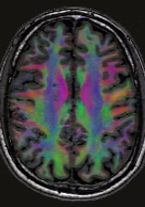
Brain
Advanced techniques such as diffusion imaging, functional MRI (fMRI), quantitative imaging and spectroscopy are used to study brain structure, connectivity, and metabolism, to understand healthy brain development and aging, and the impact of neurological conditions like stroke, multiple sclerosis, and ALS.
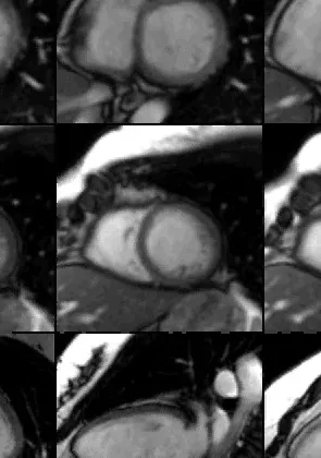
Cardiac
Specialized cardiac MRI techniques are used to assess cardiac health (e.g. in cancer patients), often utilizing MRI-compatible exercise equipment to study the effects of treatment and monitor heart function in real-time.
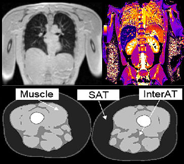
Body
Body imaging investigates lung health, body composition (muscle and fat distribution), overall body health, volumes of internal organs, and can aid in the early detection and treatment planning of diseases such as prostate cancer.
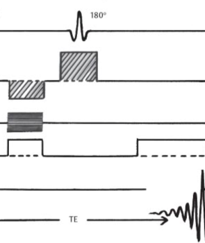
Sequence development
Creation and refinement of specialized MRI sequences tailored for specific research needs, enabling and enhancing imaging capabilities across a range of brain, cardiac, and body imaging applications.
Infrastructure and Resources
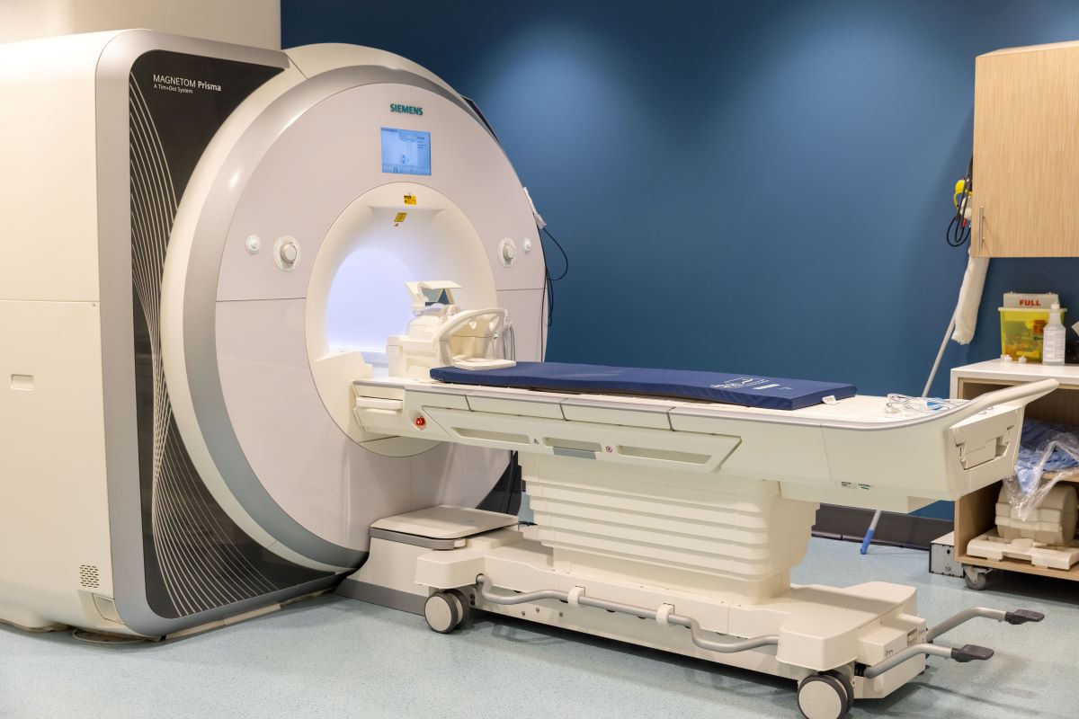
The Peter S. Allen MRI Research Centre is home to 3 fully research-dedicated MRI scanners:
3T Siemens Prisma is a state-of-the-art MRI developed by Siemens for clinical research. This system allows for development of advanced imaging techniques and offers a wide range of coils and sequences. It is equipped with a powerful gradient strength of up to 80 mT/m, enabling rapid imaging with high spatial resolution and improved signal-to-noise ratio.
Sequences available
Standard library of a Siemens medical system - Syngo XA30; addtional Siemens packages (SMS, MapIt); several "exprimental" sequences; in-house optimized sequences (heart and lung imaging, multi-nuclear imaging)
Coils available
Transmit:
- whole body (2 channel)
Receivers:
- whole body
- head/neck 20 channel
- head/neck 64 channel
- spine array 32 channel
- 2x body array 18 channel
- TxRx Knee (1Tx, 15 Rx channel) (QED)
- hand/Wrist 16 channel
- loop (3,7,11 cm)
- flexible coils (large, small) 4 channel
- 2x special purpose 4 channel
- breast coil 10+16 channel (Sentinel)
- 31P 1H TxRx surface (Rapid)
- 23Na 1H head (Rapid)
- small resonator (35mm birdcage) (Rapid)
Peripheral systems available
- MedRad power injector
- Invivo Expression patient monitor (non-invasive blood pressure, heart rate, oxygen saturation, ECG)
- projector with in-bore screen, response buttons and joystick, presentation computer (functional MRI setup)
- Elastography
- Ergostep (exercise equipment)
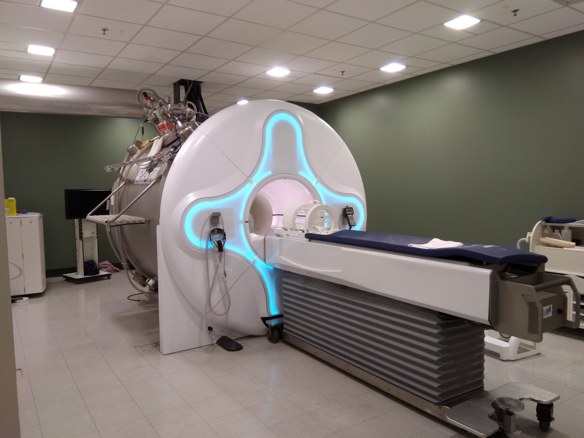
4.7T Human Research Scanner is a high-field system capable of more advanced (higher resolution) imaging and signal-to-noise than a 3T system, while avoiding the patient discomfort and technical challenges of ultra-high field systems. This system enables multi-nuclei imaging (such as sodium imaging), functional MRI, or MR spectroscopy.
4.7T system can be operated by either of the two spectrometer systems:
Legacy Varian VnmrJ (max 4 receive channels)
MR Solutions Evo (8 transmit, 32 receive channels)
Typical clinical research applications
- stroke
- MS (multiple sclerosis)
- Parkinson's
- major depression and anxiety disorders
- MedRad heart rate and and oxygen saturation monitor
- projector with in-bore screen, 32" LCD screen, response buttons, presentation computer (functional MRI setup)
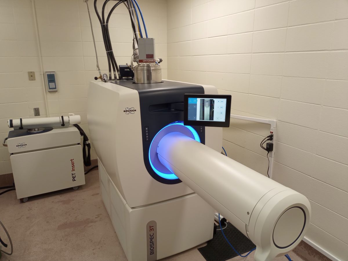
3T Bruker Pre-clinical PET/MR Scanner integrates 3T MRI technology with high-performance PET imaging, providing exceptional spatial resolution and sensitivity. Its advanced hardware enables researchers to obtain high-quality, simultaneous anatomical and functional imaging, offering powerful insights into tissue structures and metabolic processes at the pre-clinical stage.
MRI coils include volume coils with inner diameter of 23, 30, 40, 60, 72 and 82mm and 4-channel array coils for brain and cardiac imaging.
PET Insert enables simultaneous PET/MR imaging, offering excellent spatial resolution (~1.2 mm or better) and sensitivity for molecular and functional imaging studies. This allows for precise co-registration of metabolic and anatomical data.
Subject Monitoring includes respiratory gating, cardiac monitoring, and temperature regulation. This ensures optimal imaging conditions and minimizes motion artifacts, improving the accuracy of longitudinal studies.
Select Publications
T1-weighted MRI texture analysis in amyotrophic lateral sclerosis patients stratified by the D50 progression model. Parnianpour P, Steinbach R, Buchholz IJ, Grosskreutz J, Kalra S. Brain Communications. 2024 Nov 5;6(6):fcae389. doi: 10.1093/braincomms/fcae389. eCollection 2024.
Sodium MRI of the skin using a surface coil to investigate and reduce signal loss and bias. Zhu J, Beaulieu C, Damji K, Stobbe R. Magnetic Resonance in Medicine. 2024 Oct 27. doi: 10.1002/mrm.30343. Online ahead of print.
Protocol for a pilot study: Feasibility of a web-based platform to improve nutrition, mindfulness, and physical function in people living with Post COVID-19 condition (BLEND). Montes-Ibarra M, Godziuk K, Thompson RB, Chan CB, Pituskin E, Gross DP, Lam G, Schlogl M, Felipe Mota J, Paterson DI, Prado CM. Methods. 2024 Oct 9;231:186-194. doi: 10.1016/j.ymeth.2024.10.004. Online ahead of print.
Phonological, orthographic and morphological skills are related to structural properties of ventral and motor white matter pathways in skilled and impaired readers. Reed A, Huynh T, Ostevik AV, Cheema K, Sweneya S, Craig J, Cummine J. Applied Neuropsychology: Adult. 2024 Sep 17:1-12. doi: 10.1080/23279095.2024.2397036. Online ahead of print.
For a complete list of publications, see our Google Scholar page


