Alan Wilman, PhD
Contact

- Position:
- Professor and Associate Chair (Graduate Studies)
-
3-50 E University Terrace
8303 - 112 St NW
-
Edmonton
-
T6G 2T4
- This email address is being protected from spambots. You need JavaScript enabled to view it.
- 780-492-0562
- https://apps.ualberta.ca/directory/person/wilman
Additional Information
-
- Professor and Associate Chair (Graduate Studies), Dept of Biomedical Engineering, Faculty of Medicine and Engineering
- Adjunct Professor, Dept. of Physics, Faculty of ScienceEducation
BSc Physics, University of British Columbia, 1987
PhD Medical Physics, University of Alberta, 1994
Post doctorate Mayo Clinic, Rochester MN 1994-1997
My background and current work is in the physics and engineering of Magnetic Resonance Imaging (MRI). My specialty is designing new high field MRI methods to improve the value of MRI and to apply these methods to study human disease, mainly in the brain.
During my PhD, I studied with Dr. Peter S. Allen on the topic of coupled spins and spectroscopy. This work involved using spin quantum mechanics to analytically calculate the spin response of important metabolites in the human brain. My PhD gave me a strong grounding in NMR fundamentals and spectroscopy, but I wanted to also make a difference in clinical patient care. I moved to the Mayo Clinic in Minnesota to work with Dr. Stephen Riederer. At Mayo, I applied my physics understanding to discover new methods of performing MR angiography. These methods have been used on more than 20 million patients worldwide. I returned to the U of Alberta in 1997. Over the years my work at UofA has expanded from its beginnings in MR angiography and spectroscopy to include general high field brain imaging, with a current focus on iron-sensitive methods. Recent advances include new ways to perform magnetic susceptibility mapping and transverse relaxation measurements as well as new insight into multiple sclerosis grey matter iron accumulation. In all cases our goal is to make advances: new MR physics insight, new methods for the clinic, or new findings in human function and disease.
Current Research: Susceptibility and Relaxation and Iron-Sensitive MRI

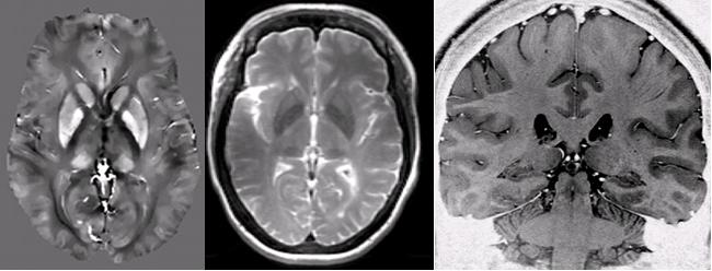
At the University of Alberta we have a 100% research 4.7T human MRI system and a 3T Prisma. These high field instruments offer unique sensitivity to iron through susceptibility and relaxation effects. Iron may be an early marker of neurodegeneration in the brain, as well as being an indicator of changes in deoxygenation and iron storage. It is an extremely important element in the human brain and we are lucky to be sensitized to its measurement when using high field MRI. Our work is finding that iron may be a biomarker of disease state, particularly for multiple sclerosis. More generally, we exploit MRI physics and engineering to enhance imaging methods or "pulse sequences" in new ways to gain more value from MRI. We are interested in susceptibility, structural and relaxation methods, as well as blood vessel imaging and spectroscopy. See my publication section for detailed reading.
A general comment is we want to bring about revolutions in the use of MRI by exploring untapped research areas. We generally do modelling in MATLAB and then apply our advances to normal volunteers and to specific diseases where the new methods can make a difference. One of the strengths of the program is the wide range of collaborating physicians in neurological and psychiatric diseases including depression, Parkinson's disease, multiple sclerosis, stroke, blood vessel disease and dementia. Once we develop new methods, we can apply them to a wide range of diseases within our MRI center with their help.
Another important aspect is that we own all our MRI machines and thus they are available for research all day long. Most students are thus able to complete all scanning needs in daytime hours and lead normal lives. Lastly from an international perspective, the ranking of the University can also be important, where University of Alberta is typically the 4th ranked school in Canada.
Graduate Students
As a general rule, potential graduate students should start by examining the professor's track record and ask four key questions: Are the grad students getting first author publications? Am I interested in the professor's work? Do I have the right background to succeed? Are there adequate resources? Students working with me generally have a strong background in electrical engineering, physics, or engineering physics. Students can enter either the Biomedical Engineering Department or the Physics Department. Students learn how to control (program) the MRI scanners and often create new ways of performing or processing MRI. We then apply our methods to patient studies. Recent advances have included new analysis methods for transverse relaxometry, new ways of performing susceptibility mapping and demonstration of iron as a biomarker in multiple sclerosis. Some advances can make a true difference in diagnostic imaging, while others provide new avenues for brain research. Graduating students typically find places in industry or in MRI research hospital and academic environments. Some also enter medical school. MRI remains a growing field with good job opportunities.
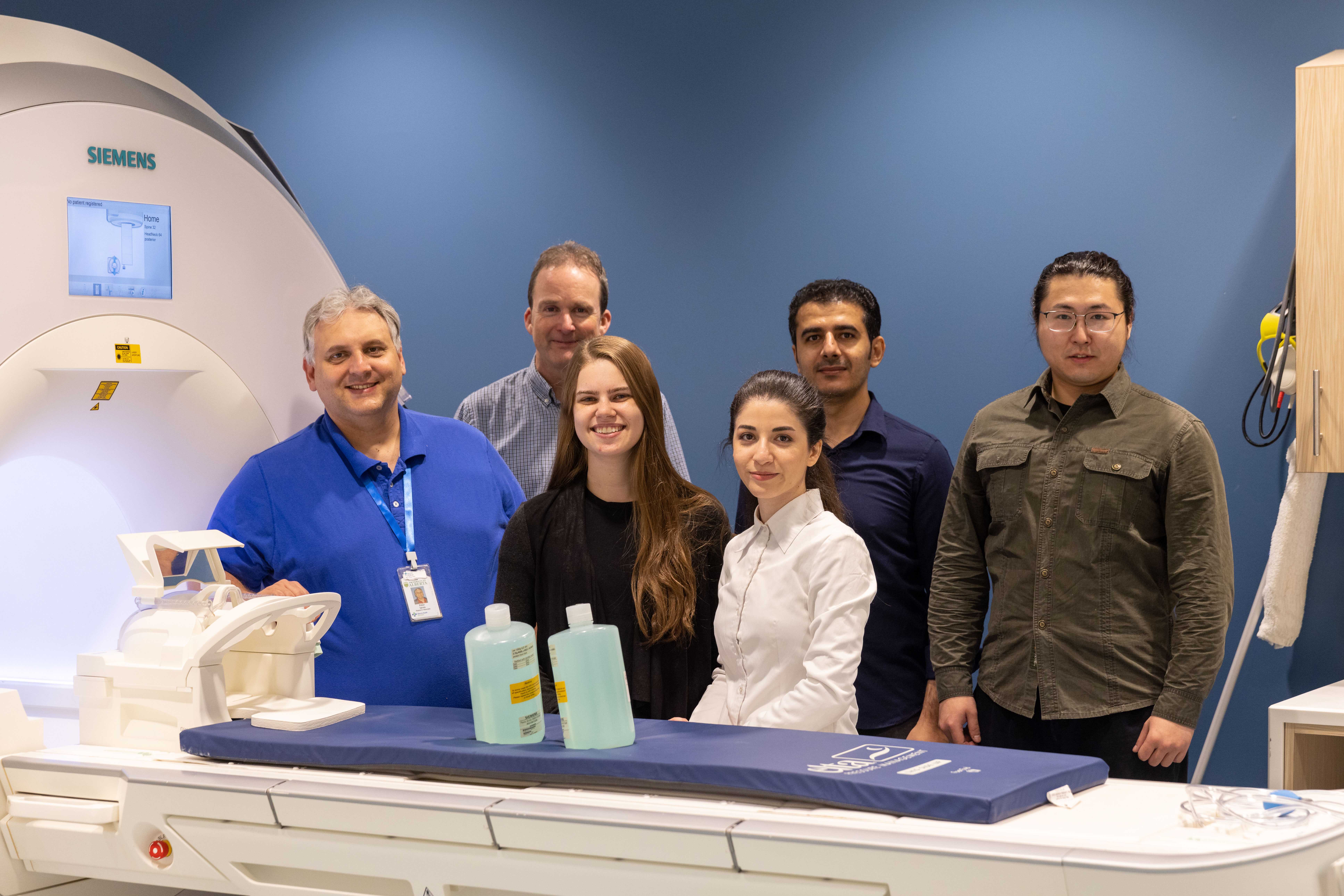
Typically I have about 4 graduate students. I like to keep the group relatively small to maximize interaction and learning. Also this way projects are not watered down and a single grad student gets the chance to make major innovations. Our department in total has approximately 15 MRI graduate students and four MRI professors, as well as technical staff which make it an excellent center for making advances in MRI.
To learn more about graduate studies in the BME department click here.
Publications
Most of my publications can be found at www.pubmed.org just type in Wilman A, and you will get an up-to-date list. Most publications can be downloaded, but also feel free to contact me to learn more about my research.
You can also review a list of my publications - with trainees underlined.
Research Support
Four agencies have been extremely helpful to me in providing financial support:
Canadian Institutes of Health Research (CIHR)
Natural Science and Engineering Research Council of Canada (NSERC)
Multiple Sclerosis Society of Canada
The University Hospital Foundation
Also the Canadian Foundation for Innovation (CFI) helped establish our MRI centre in 1999 and helped us renew it in 2014.


